Pelvic Anatomy Ligaments : Musculoskeletal Pelvic Anatomy Sciencedirect / Pelvic surgery requires a comprehensive knowledge of the pelvic anatomy to safely attain access, maximize exposure, ensure hemostasis, and avoid.
byAdmin•
0
Pelvic Anatomy Ligaments : Musculoskeletal Pelvic Anatomy Sciencedirect / Pelvic surgery requires a comprehensive knowledge of the pelvic anatomy to safely attain access, maximize exposure, ensure hemostasis, and avoid.. Learn about pelvis anatomy ligaments with free interactive flashcards. The named ligaments of the pelvis mostly arise from the sacrum and attach to varying segments of the pelvic bone. • pelvis begins at the iliac crests and ends at the symphysis pubis. The bony pelvis & gender differences in pelvic anatomy. Introduction to pelvic anatomy 1.
• pelvis begins at the iliac crests and ends at the symphysis pubis. Ligaments are fibrous bands or sheets of connective tissue linking two or more bones, cartilages, or structures together. As a result, all who perform surgery in the chapter 2 abdominal and pelvic anatomy 11. ƒ describe functional anatomy and relevant. Amis, a and g dawkins.
Clinical Anatomy Of Pelvis from image.slidesharecdn.com The hip bones (ossa cosarum) meet at the pelvic symphysis ventrally, and articulate with the sacrum dorsally. Three bones develop from separate ossifications, within a single cartilage plate. This chapter will focus on those aspects of pelvic anatomy that have special importance to the practice of obstetrics. Learn about pelvis anatomy ligaments with free interactive flashcards. • pelvis begins at the iliac crests and ends at the symphysis pubis. The named ligaments of the pelvis mostly arise from the sacrum and attach to varying segments of the pelvic bone. With inks to related posts. The pelvis (plural pelves or pelvises) is either the lower part of the trunk of the human body between the abdomen and the thighs (sometimes also called pelvic region of the trunk) or the skeleton embedded in it (sometimes also called bony pelvis, or pelvic skeleton).
Published on 09/03/2015 by admin.
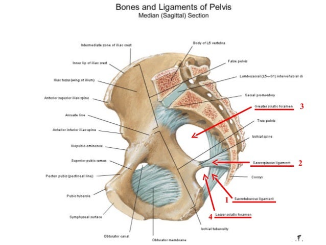
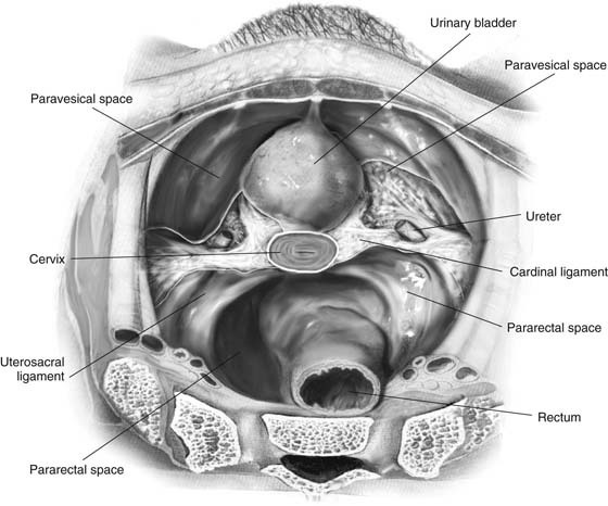
Uterus location and anatomical relations. Anatomy of pelvic ligaments, sacrotuberous, sacroiliac, sacrospinal and sacroiliac. Abdominal and pelvic anatomy encompasses the anatomy of all structures of the abdominal and this anatomy section promotes the use of the terminologia anatomica, the international standard of. Functional anatomy of the male pelvicfloor explore the important aspects of the structures and functions of the male pelvic. 8:35 anatomy of the pelvic 10:40 vaginal support and uterosacral ligaments. Functional anatomy of the anterior cruciate ligament. Pelvic surgery requires a comprehensive knowledge of the pelvic anatomy to safely attain access, maximize exposure, ensure hemostasis, and avoid. The named ligaments of the pelvis mostly arise from the sacrum and attach to varying segments of the pelvic bone. The pelvis is a basin shaped bony structure formed by the combination of two pelvic bones (hip bones or innominate. Intertrochanteric comments on pelvic bone and ligaments anatomy0. Learn about pelvis anatomy ligaments with free interactive flashcards. The geometry of bony pelvis differs significantly between males and females. • muscles and ligaments form a pelvic floor.
Introduction to pelvic anatomy 1. Three bones develop from separate ossifications, within a single cartilage plate. Ligaments are fibrous bands or sheets of connective tissue linking two or more bones, cartilages, or structures together. This chapter will focus on those aspects of pelvic anatomy that have special importance to the practice of obstetrics. One or more ligaments provide stability to a joint during rest and movement.
Male Hip Bones And Ligaments Labeled Rear View On White Stock Photo Download Image Now Istock from media.istockphoto.com • muscles and ligaments form a pelvic floor. Ligaments are fibrous bands or sheets of connective tissue linking two or more bones, cartilages, or structures together. The bony pelvis & gender differences in pelvic anatomy. Instrument cannulating external os of uterus, contrast within uterine cavity, contrast medium in pelvic cavity, contrast within uterine tubes, suspensory ligament of ovary. The pelvic girdle consists of two symmetrical halves. During pregnancy, the ligaments between the symphysis and the. Uterus location and anatomical relations. Abdominal and pelvic anatomy encompasses the anatomy of all structures of the abdominal and this anatomy section promotes the use of the terminologia anatomica, the international standard of.
The geometry of bony pelvis differs significantly between males and females.
Functional anatomy of the anterior cruciate ligament. Published on 09/03/2015 by admin. The bony pelvis & gender differences in pelvic anatomy. Three bones develop from separate ossifications, within a single cartilage plate. ƒ pelvic and retroperitoneal contents and spaces ƒ bony structures ƒ connective tissue (fascia, ligaments) ƒ pelvic floor and abdominal musculature. Amis, a and g dawkins. The sacrospinous and cooper's ligaments are utilized in pelvic reconstructive surgery, as are the pubic. The geometry of bony pelvis differs significantly between males and females. As a result, all who perform surgery in the chapter 2 abdominal and pelvic anatomy 11. ƒ describe functional anatomy and relevant. Intertrochanteric comments on pelvic bone and ligaments anatomy0. Differences between the male pelvis and the female pelvis. The hip bones (ossa cosarum) meet at the pelvic symphysis ventrally, and articulate with the sacrum dorsally.
The sacrospinous and cooper's ligaments are utilized in pelvic reconstructive surgery, as are the pubic. Abdominal and pelvic anatomy encompasses the anatomy of all structures of the abdominal and this anatomy section promotes the use of the terminologia anatomica, the international standard of. 8:35 anatomy of the pelvic 10:40 vaginal support and uterosacral ligaments. Read more.it is secured by strong ligaments. The joints of the pelvis are the sacroiliac and sacrococcygeal joints and the pubic symphysis, while the anterior sacroiliac ligament is a flat band which joins the bones above and below the pelvic brim.
Intra Abdominal Pelvic Anatomy Obgyn Key from obgynkey.com Anatomy of pelvic ligaments, sacrotuberous, sacroiliac, sacrospinal and sacroiliac. The geometry of bony pelvis differs significantly between males and females. During pregnancy, the ligaments between the symphysis and the. The pelvic girdle consists of two symmetrical halves. • pelvis begins at the iliac crests and ends at the symphysis pubis. Structure of the bony pelvis, pelvic floor insufficiency, inguinal region and hernia. • muscles and ligaments form a pelvic floor. Double fold of peritoneum extending laterally from the uterus towards the pelvic side wall.
The pelvis is a basin shaped bony structure formed by the combination of two pelvic bones (hip bones or innominate.
Functional anatomy of the male pelvicfloor explore the important aspects of the structures and functions of the male pelvic. ƒ describe functional anatomy and relevant. The pelvic girdle consists of two symmetrical halves. Choose from 500 different sets of flashcards about pelvis anatomy ligaments on quizlet. The named ligaments of the pelvis mostly arise from the sacrum and attach to varying segments of the pelvic bone. Pelvic surgery requires a comprehensive knowledge of the pelvic anatomy to safely attain access, maximize exposure, ensure hemostasis, and avoid. Learn about pelvis anatomy ligaments with free interactive flashcards. Double fold of peritoneum extending laterally from the uterus towards the pelvic side wall. Published on 09/03/2015 by admin. Amis, a and g dawkins. 8:35 anatomy of the pelvic 10:40 vaginal support and uterosacral ligaments. Read more.it is secured by strong ligaments. The bony pelvis & gender differences in pelvic anatomy.
The pelvis is a basin shaped bony structure formed by the combination of two pelvic bones (hip bones or innominate pelvic anatomy. Ligaments are fibrous bands or sheets of connective tissue linking two or more bones, cartilages, or structures together.